
Knee Arthroscopy
Knee-related advancement shown in its key-hole surgery. Each surgery done with the help of video-camera is known as knee arthroscopy.
The main attraction is to fix up a surgery with a small camera called an Arthroscope inserted into the joint. Part for patient to get it done with a small cut of 1 cm.
The main surgery is done looking at the monitor of the camera and the instruments inside the joint. This enables the surgeon to operate through a small entrance. Advantages: being quick and early recovery compared to bone surgery.
While the knee is the joint most often used and operated today, these arthroscopic procedures such as the shoulder, elbow, ankle, hip, and wrist can also be done using this instrument.
How is the knee form inside?
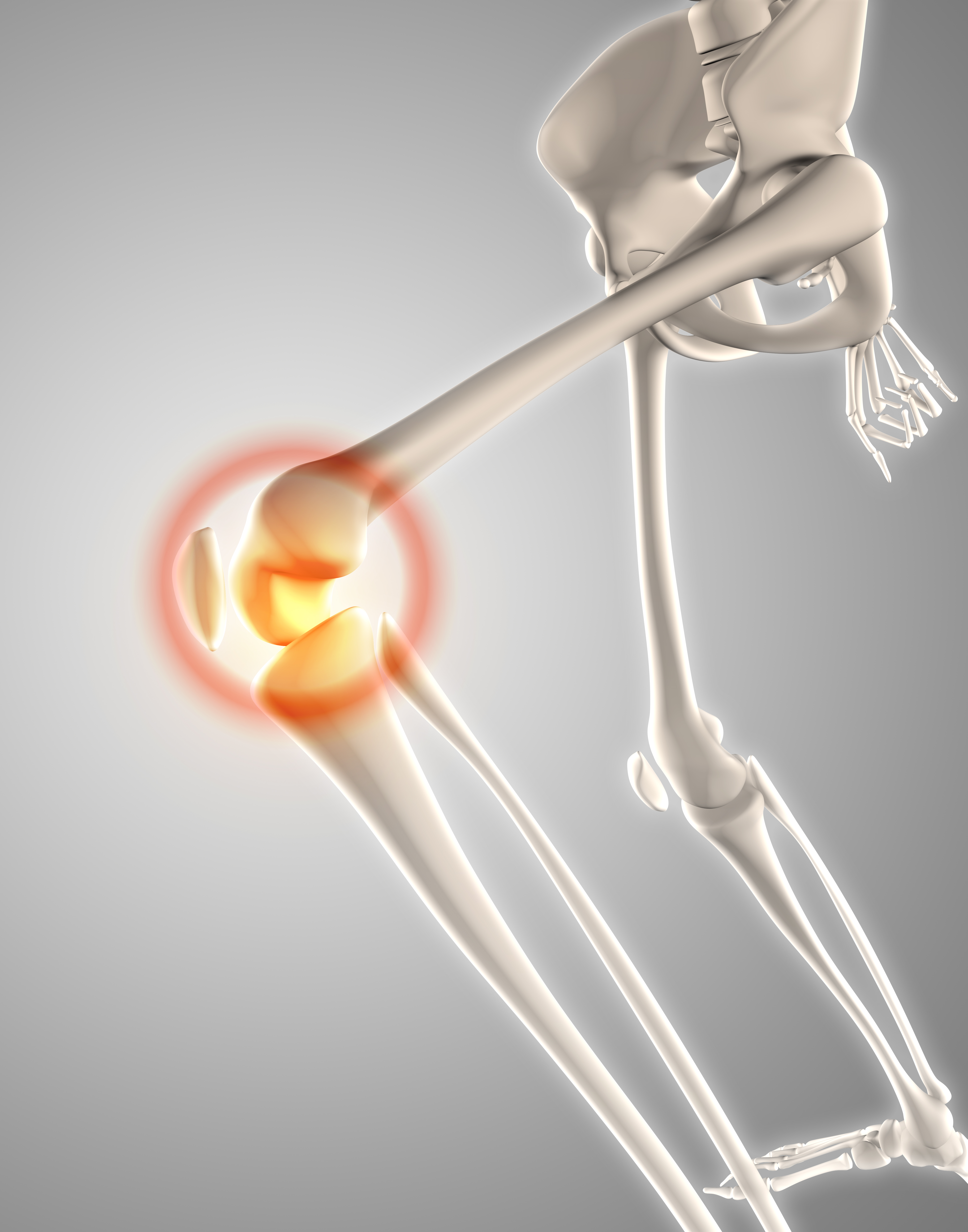
Parts of the joint include:
Ligaments: There are two ligaments in the joint and two ligaments outside the joint.
Firstly, the inside the knot ligaments: Depending on the inside foundation they are named as Anterior Cruciate (ACL) and Posterior Cruciate Ligament (PCL). They cross each other in the joint. These ligaments prevent the thigh bone from sliding back and forth.
Two ligaments are outside the joints. There are Collateral Ligaments: Medial Collateral on the medial side and Lateral Collateral on the lateral side.
Meniscus: These are two washer-shaped rubbing pieces in between the bones which reduce the friction of the joint. They are important in cushion and load-bearing function as well as stability of the joint.
Articular Cartilage: The bone ends are covered with smooth surface called cartilage. This allows smooth sliding of the joint. It is between cartilage and cartilage.
Indication of knee arthroscopy surgery:
ACL Rupture
ACL is a common injury where the ligament within the knee breaks. Arthroscopy is the standard of care to reconstruct the ligament.
Meniscal Tear
A tear in the cushion (meniscus) of the knee can lead to pain and locking. Today we try to preserve and repair the meniscus surface wherever possible.
Patellar Dislocation
The kneecap (patella) slips out of its groove. Arthroscopic lateral release or ligament reconstruction may be needed.
Other indications include loose bodies, synovitis, and diagnostic biopsy in complex cases.